Understanding Autism and the Brain
To comprehend the intricate relationship between autism and the brain, it is essential to explore the scientific findings that shed light on this enigma. Two crucial aspects to consider are the link between glutamate and autism, as well as multisensory integration in individuals with autism.
Linking Glutamate to Autism
Glutamate, a neurotransmitter involved in brain signaling, has been implicated in various mental health disorders, including autism, depression, and schizophrenia. Research suggests that problems in the production or utilization of glutamate may contribute to the development of autism.
While the precise mechanisms are still being studied, alterations in glutamate levels and receptor function have been observed in individuals with autism. These disruptions in glutamate signaling may impact neural circuits involved in social communication, cognition, and sensory processing, contributing to the characteristic features of autism spectrum disorder (ASD).
Multisensory Integration in Autism
Multisensory integration refers to the brain’s ability to combine information from various sensory modalities, such as sight, sound, touch, and smell, to form a cohesive perception of the world. In individuals with autism, difficulties in multisensory integration have been reported.
Impairments in multisensory integration can manifest in deficits in imitation, language development, emotion recognition, and social bonding. Autistic individuals may struggle to integrate and interpret incoming sensory stimuli, leading to challenges in understanding social cues and engaging in reciprocal interactions.
By investigating the link between glutamate and autism, as well as the complexities of multisensory integration in individuals with autism, researchers aim to unravel the underlying mechanisms of this neurodevelopmental condition. These insights can potentially pave the way for innovative interventions and therapies to improve the lives of individuals with autism.
Sensory Processing in Autism
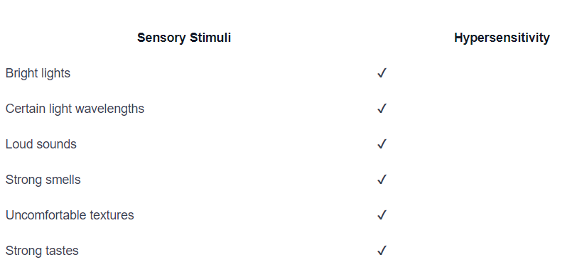
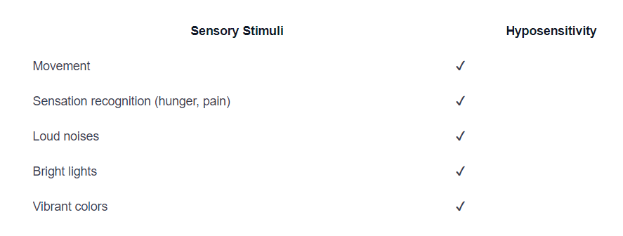
One of the defining features of autism spectrum disorder (ASD) is the presence of sensory issues. Sensory processing difficulties are common among individuals with autism and can significantly impact their experiences and behaviors. These issues encompass both hypersensitivity (over-responsiveness) and hyposensitivity (under-responsiveness) to various stimuli, often exhibiting a combination of both.
Hypersensitivity in Autism
Hypersensitivity refers to an increased sensitivity or over-responsiveness to sensory stimuli. Many individuals with autism experience hypersensitivity to bright lights, certain light wavelengths, sounds, smells, textures, and tastes. These heightened sensitivities can lead to sensory avoidance behaviors, such as pulling away from physical touch or covering ears to escape loud sounds.

Hyposensitivity in Autism
Hyposensitivity, on the other hand, refers to a reduced sensitivity or under-responsiveness to sensory stimuli. Individuals with autism may exhibit hyposensitivity, resulting in behaviors such as a constant need for movement, difficulty recognizing sensations like hunger or pain, and attraction to loud noises, bright lights, and vibrant colors. These individuals may engage in sensory seeking activities to fulfill their sensory needs.

Sensory Overload and Autism
Sensory overload occurs when intense sensory stimuli overwhelm an individual’s ability to cope, leading to feelings of intense anxiety, a need to escape, or difficulty communicating. During sensory overload, the brain may prioritize sensory processing over other functions such as speech, decision-making, and information processing. This can result in temporary shutdowns or meltdowns as a way to cope with the overwhelming sensory experiences.
Understanding and accommodating the sensory needs of individuals with autism is crucial for enhancing their comfort and increasing opportunities for learning, socializing, and participating in the community. By recognizing and respecting their sensory sensitivities, we can create supportive environments that promote well-being and improve their overall quality of life [3].
Brain Structure in Autism
The study of brain structure in individuals with autism has provided valuable insights into the neurodevelopmental differences associated with the condition. In this section, we will explore three key areas of brain structure that have been found to be affected in autism: the enlarged hippocampus, cerebellum differences, and the corpus callosum.
Enlarged Hippocampus in Autism
Research has shown that children and adolescents with autism often have an enlarged hippocampus, a region of the brain involved in memory and learning. However, it is still unclear whether this difference persists into adolescence and adulthood [4]. The significance of this enlargement and its relationship to the cognitive and behavioral characteristics of autism are still being studied.
Cerebellum Differences in Autism
Another area of interest in autism research is the cerebellum. Autistic individuals have been found to have decreased amounts of brain tissue in certain parts of the cerebellum. The cerebellum plays a crucial role in motor coordination, but it is also involved in cognition and social interaction. The impact of these cerebellar differences on the symptoms and characteristics of autism is an ongoing area of investigation.
Corpus Callosum and Autism
The corpus callosum, a white matter tract that connects the two hemispheres of the brain, has also been implicated in autism. Research suggests that individuals who lack all or part of the corpus callosum have an increased likelihood of being autistic or exhibiting traits associated with autism. This supports the connectivity theory of autism, which suggests that alterations in brain connectivity may contribute to the condition.
Understanding the structural differences in the brains of individuals with autism provides valuable insights into the underlying mechanisms of the condition. Further research is needed to fully elucidate the relationship between these structural differences and the cognitive, behavioral, and social characteristics of autism. By unraveling the enigma of autism’s impact on the brain, we can continue to advance our understanding of this complex condition and develop more effective strategies for support and intervention.
White Matter Changes in Autism
In individuals with autism, white matter, the bundles of long neuron fibers that connect different brain regions, undergo significant alterations. These changes in white matter structure have been observed in toddlers, preschoolers, and adolescents with autism, indicating widespread differences throughout the brain.
White Matter Alterations in Toddlers
The structural differences in white matter tracts have been found in autistic toddlers. Diffusion MRI studies have revealed deviations in white matter organization, suggesting disruptions in the connectivity between brain regions at an early age. These alterations in white matter may contribute to the connectivity issues seen in individuals with autism.
Over-Connectivity in Autism
A common neuroanatomical theme in autism is over-connectivity in closely related areas and decreased connectivity in longer circuitry requiring large-scale integration. Studies have shown disordered development of grey and white matter in autistic individuals, particularly in the frontal and temporal cortices. This disordered development includes a selective increase in late-developing white matter and narrow mini-columns in the frontal and temporal cortex, which have been associated with early accelerated postnatal head growth.
The observed over-connectivity within closely related brain areas and decreased connectivity in longer circuits may contribute to the characteristic cognitive and behavioral differences seen in individuals with autism. Understanding these white matter alterations is crucial for unraveling the complexities of autism and its impact on brain connectivity.
By investigating white matter changes in autism, researchers aim to gain further insights into the underlying mechanisms of this neurodevelopmental disorder. The findings contribute to a better understanding of how alterations in white matter structure may influence brain connectivity and the cognitive functions associated with autism.
Social Cognition in Autism
Autism is often characterized by deficits in social cognition, which refer to difficulties in understanding and interpreting social cues and interactions. These deficits can have a significant impact on the ability of individuals with autism to navigate social situations effectively. In this section, we will explore two key aspects of social cognition in autism: social brain network dysfunction and theory of mind deficits.
Social Brain Network Dysfunction
Research has identified social brain network dysfunction as a key contributor to social cognition deficits in individuals with autism. Social brain network dysfunction refers to reduced synchronization between specific brain regions during various social and emotional tasks, leading to impaired social cognition abilities. This suggests that autism can be considered a “neural connectivity disorder.”
The social brain network encompasses several brain regions, including the fusiform face area, inferior frontal gyrus, posterior superior temporal sulcus, superior frontal gyrus, and the amygdala. Dysfunction in these regions can contribute to difficulties in recognizing and interpreting facial expressions, understanding emotions, and processing social information.
Impaired interactions with primary caregivers during critical developmental periods can disrupt the normal development of social cognition, leading to deficits that impact language development and behavioral functioning later in life. Understanding the neural underpinnings of social brain network dysfunction is crucial for developing interventions and therapies that can help individuals with autism improve their social cognition abilities.
Theory of Mind Deficits
Theory of Mind (TOM) refers to the ability to understand and infer the mental states and perspectives of others. Individuals with autism often experience deficits in theory of mind, which can impact their ability to engage in social interactions and understand the thoughts, feelings, and intentions of others.
Studies have shown that TOM deficits in autism are associated with abnormal patterns of brain activation and connectivity. Specifically, hypoactivation has been observed in brain regions such as the superior temporal gyrus, superior temporal sulcus, and basal temporal areas, which are involved in social cognition and understanding others’ mental states. On the other hand, hyperactivation has been observed in Brodmann’s area 9/10, which is associated with cognitive control and executive functions.
These abnormal patterns of brain activation suggest that individuals with autism may struggle to put themselves in others’ shoes and understand their perspectives. This can lead to difficulties in empathizing with others and comprehending social situations accurately.
Understanding the social cognition deficits, including social brain network dysfunction and theory of mind deficits, is essential for developing targeted interventions and therapies that can help individuals with autism improve their social skills, enhance their interactions with others, and navigate social environments more effectively. Ongoing research in this area holds promise for developing effective strategies to support individuals with autism in their social development.
Neuroanatomical Differences in ASD
Understanding the neuroanatomical differences in individuals with Autism Spectrum Disorder (ASD) provides valuable insights into the underlying mechanisms of the condition. Extensive research has identified variations in brain structure and function that contribute to the unique characteristics of ASD. In this section, we will explore three key neuroanatomical differences: frontal cortex abnormalities, cerebellum involvement, and amygdala changes.
Frontal Cortex Abnormalities
The frontal cortex, responsible for executive functions and social cognition, exhibits abnormalities in individuals with ASD. Studies have shown differences in the growth and organization of the frontal cortex in those with ASD compared to neurotypical individuals [6]. Longitudinal research has revealed that the frontal cortex initially undergoes overgrowth in early childhood, followed by a reduction in size later on. These alterations in regional identity and neural stem cell expansion may contribute to the increased brain size observed in ASD.
Cerebellum and ASD
The cerebellum, a structure involved in coordination and cognitive functions, has also been implicated in ASD. Studies have identified abnormalities in the cerebellar vermis and alterations in the number and size of Purkinje cells in individuals with ASD. These findings suggest that the developmental origin and underlying causes of cerebellar abnormalities play a critical role in the behavioral consequences observed in ASD.
Amygdala Changes in ASD
The amygdala, a structure involved in emotional processing, has been found to undergo growth pattern alterations in individuals with ASD. Research has shown increased amygdala volume in young children with ASD, which correlates with social and communication deficits. The precise role of the amygdala in ASD symptoms, such as increased anxiety and deficits in social behavior, is still a topic of ongoing debate [6].
Understanding these neuroanatomical differences provides valuable insights into the underlying mechanisms of ASD. Abnormalities in the frontal cortex, cerebellum, and amygdala contribute to the unique characteristics and challenges faced by individuals with ASD. Continued research into these neuroanatomical differences will help further unravel the enigma of ASD and pave the way for targeted interventions and treatments.
References
- https://my.clevelandclinic.org/health/articles/22839-glutamate
- https://www.ncbi.nlm.nih.gov/pmc/articles/PMC6987885/
- https://www.autismspeaks.org/sensory-issues
- https://www.thetransmitter.org/spectrum/brain-structure-changes-in-autism-explained
- https://www.ncbi.nlm.nih.gov/pmc/articles/PMC4117155/
- https://www.ncbi.nlm.nih.gov/pmc/articles/PMC5192959/

 We've just released an article!
Check out our blog!


